Circulating tumor cells (CTCs) are a rare type of cancer cell that are found in the blood stream of patients with localized tumors. Successful separation of CTCs from blood could serve as a liquid biopsy to help diagnose cancer and monitor treatment progress. A deeper understanding of CTCs could also lead to a better understanding of the most deadly cancer process: metastasis, where cancer cells leave established tumors and migrate to other locations in the body.
Currently, CTC separation methods rely on features that distinguish CTCs from other cells—antibodies that stick to them, cell size, deformability, or even electrical properties. Scientists have also explored using sound waves to separate CTCs. Acoustic-based separation provides excellent biocompatibility and safety; it preserves the viability, function, phenotype, and genotype of cells. It also allow cells to be separated without modification. As a result, sound-based separation methods enable CTCs to be maintained in their native state throughout the separation process while avoiding invasive biopsies.
Unfortunately, previous sound-based separations technologies haven't managed to separate CTCs from clinical samples due to insufficient throughput and long-term operational instability. Recently, a team of scientists has developed an acoustic-based microfluidic device that separates CTCs from peripheral blood samples of cancer patients in a high-throughput manner. This method relies on something called tilted-angle standing surface sound waves. These standing waves contain points that “stand still,” called nodes, around which the wave oscillates.
In this design, a microfluidic channel is surrounded by interdigitated transducers, which convert power into sound waves. A series of pressure nodes and antinodes are established in the microfluidic channel at an angle that is tilted relative to the fluid flow direction.
When cells flow through the channel, they pass multiple regions of paired pressure nodes and antinodes. In each of these regions, the cell experiences a different force caused by the acoustic radiation, leading to slightly different trajectory paths. As the cells pass through more and more of these features, they amplify the differences in trajectory angle, which results in cell path differences that are several orders of magnitude larger than the acoustic wavelength. Without this amplification process, previous systems have had separation distances limited to a quarter of the wavelength of the sound.
The team investigated the effect of a few key parameters on the success of this process using a simulation. They were able to optimize the flow rate, power input, tilt angle, and the length at which the acoustic wave is applied by the transducers. They found that as flow rate increases, the optimal tilt angle decreases because there are longer traveling times between pressure antinodes and nodes. The longer it takes cells to travel through the acoustic wave field, the longer the separation distance will be. At the same time, applying sound over longer distances requires less energy, decreasing the primary wave force. These two competing factors influence the optimization of the acoustic wave apparatus.
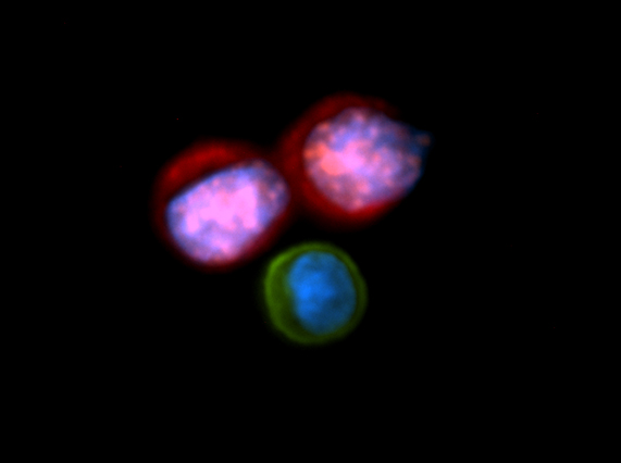
The scientists also validated their system experimentally by separating a variety of cancer cells from white blood cells. They used lithium niobate as a piezoelectric substrate to form the transducers. The microfluidic channel was composed of a soft polymer, polydimethylsiloxane.
They found that using predetermined settings with high power-inputs can result in greater than 83 percent cancer cell recovery for a variety of cancer cells. Each cancer cell population has slightly different physical properties to which the system could be tailored, but these results demonstrate that the parameters do not necessarily need to be so finely tuned for each cell population. CTCs separated using this method were viable, undamaged, and could be subsequently cultured.
This sound-based separations technique may ultimately help us isolate sufficient CTCs to help inform cancer diagnosis and treatment. However, many more studies must be performed assessing its limitations before this team can implement their device in the field. Nevertheless, some of the researchers involved have filed for a patent, suggesting they're certainly going to try.
PNAS, 2014. DOI: 10.1073/pnas.1504484112 (About DOIs).
reader comments
28